유효성 평가
의약품, 식품첨가물, 건강기능식품 등의 생리학적 작용과 효능 판단 시험입니다.

안과 시험
Ocular Disease model
Laser induced CNV model(Wet AMD model)
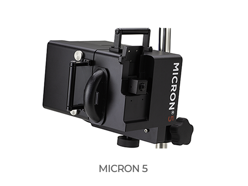
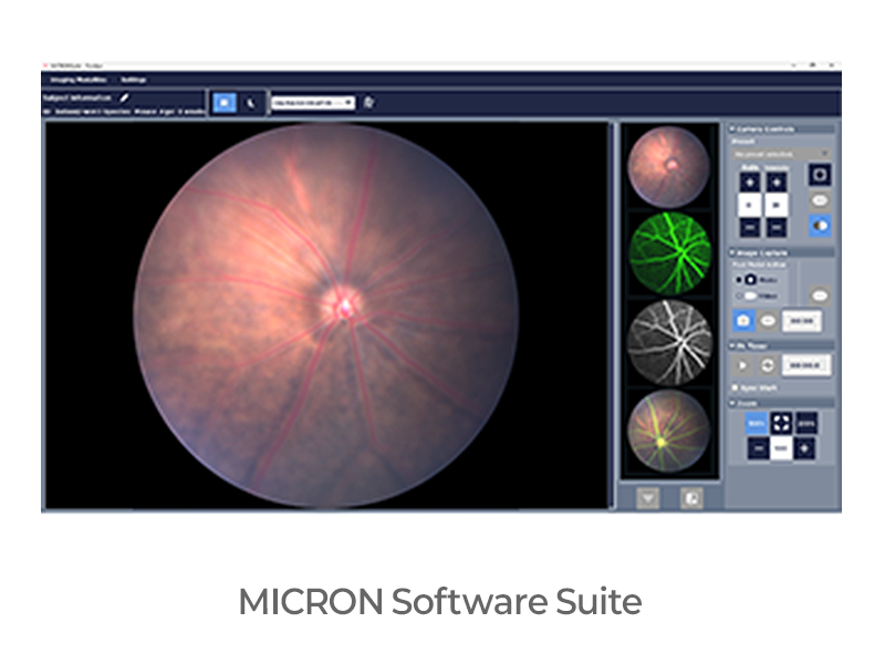
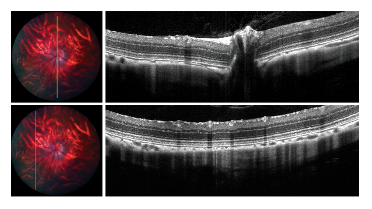
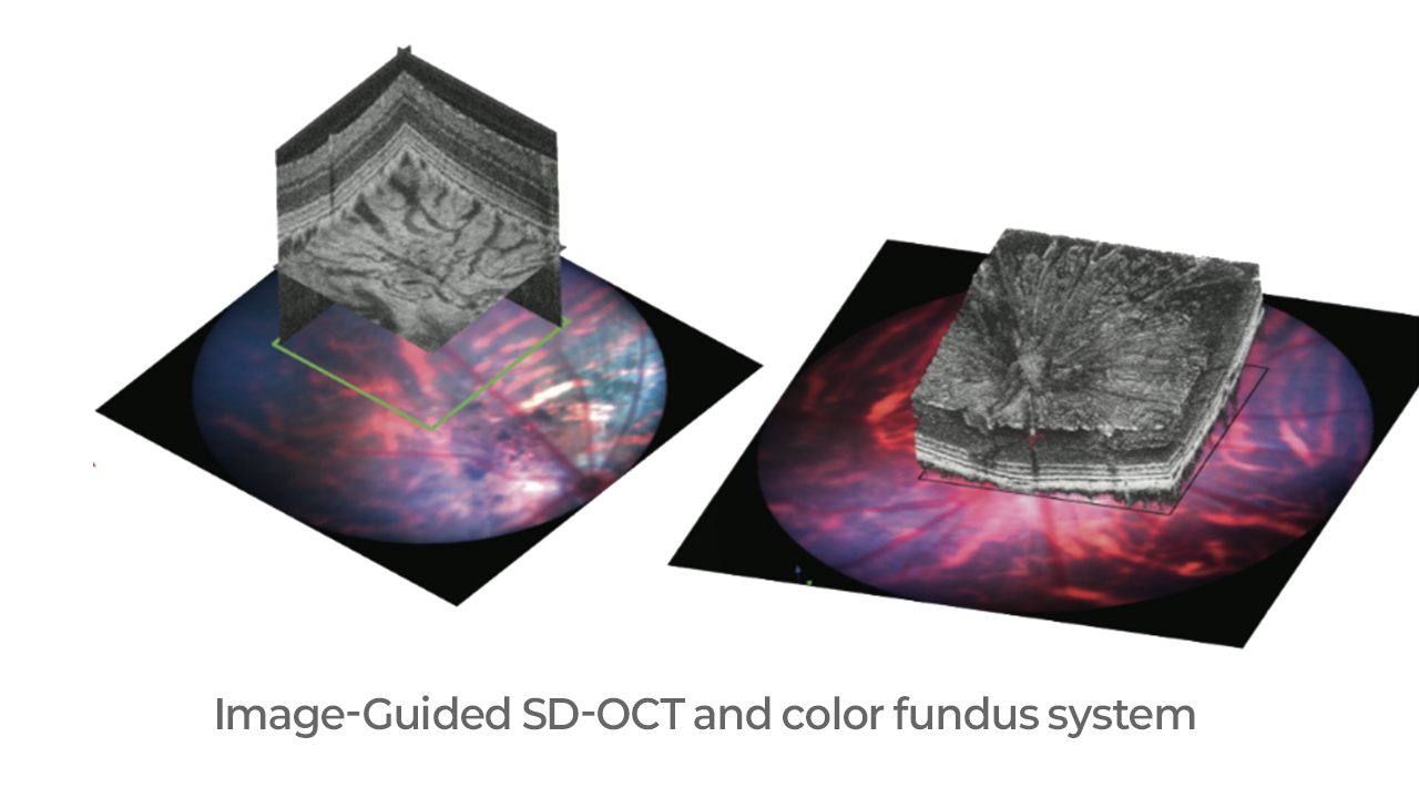
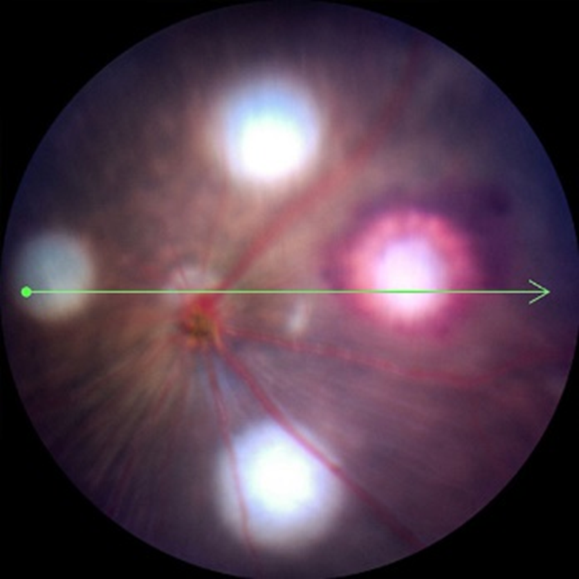
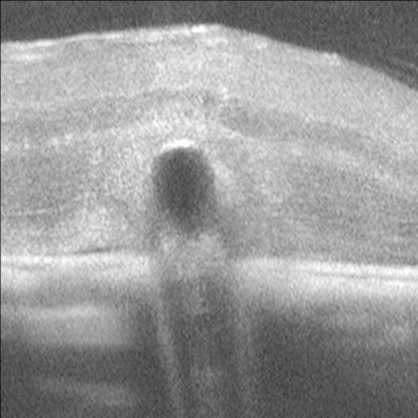
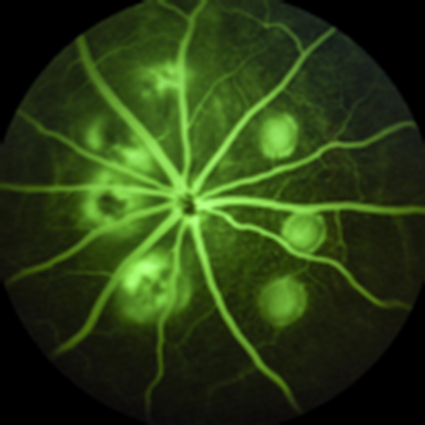
- Image-guided OCT / funduscopy
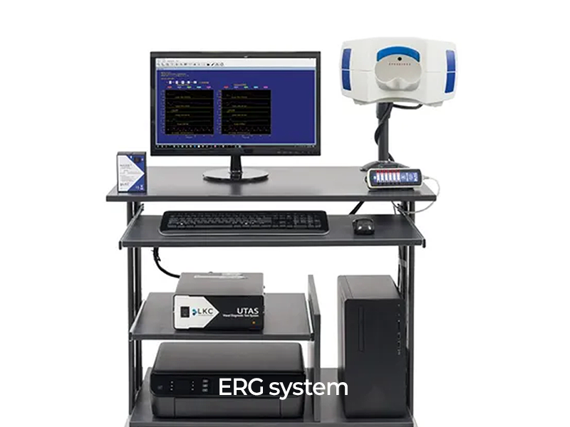
- Electroretinography
- Immunohistochemistry
- Further optional evaluation
Blue LED induced damage(Dry AMD model)
- Image-guided OCT / funduscopy
- Electroretinography
- Immunohistochemistry
- Further optional evaluation
Dry AMD rat model
- Cytokine: TNF-α, IL-1β, IL-6 (serum ELISA)
- Antioxidant factor : TAC, SOD, CAT, GSH-Px (serum ELISA)
- Anti-inflammatory factor : MDA (serum ELISA)
- Retinal damage : NF-kB, Nrf-2, HO-1 (Retina qPCR)
- Histology : ONL thickness, ONL nuclear number, Photoreceptor thickness
Sodium Iodate-induced RD model
- Electroretinography
- Image-guided OCT / funduscopy
- Immunohistochemistry
MNU-induced RD model
- Electroretinography
- Image-guided OCT / funduscopy
- Immunohistochemistry
Humanized RS1-mut mouse model
Diabetic Retinopathy model
- Image-guided OCT / funduscopy
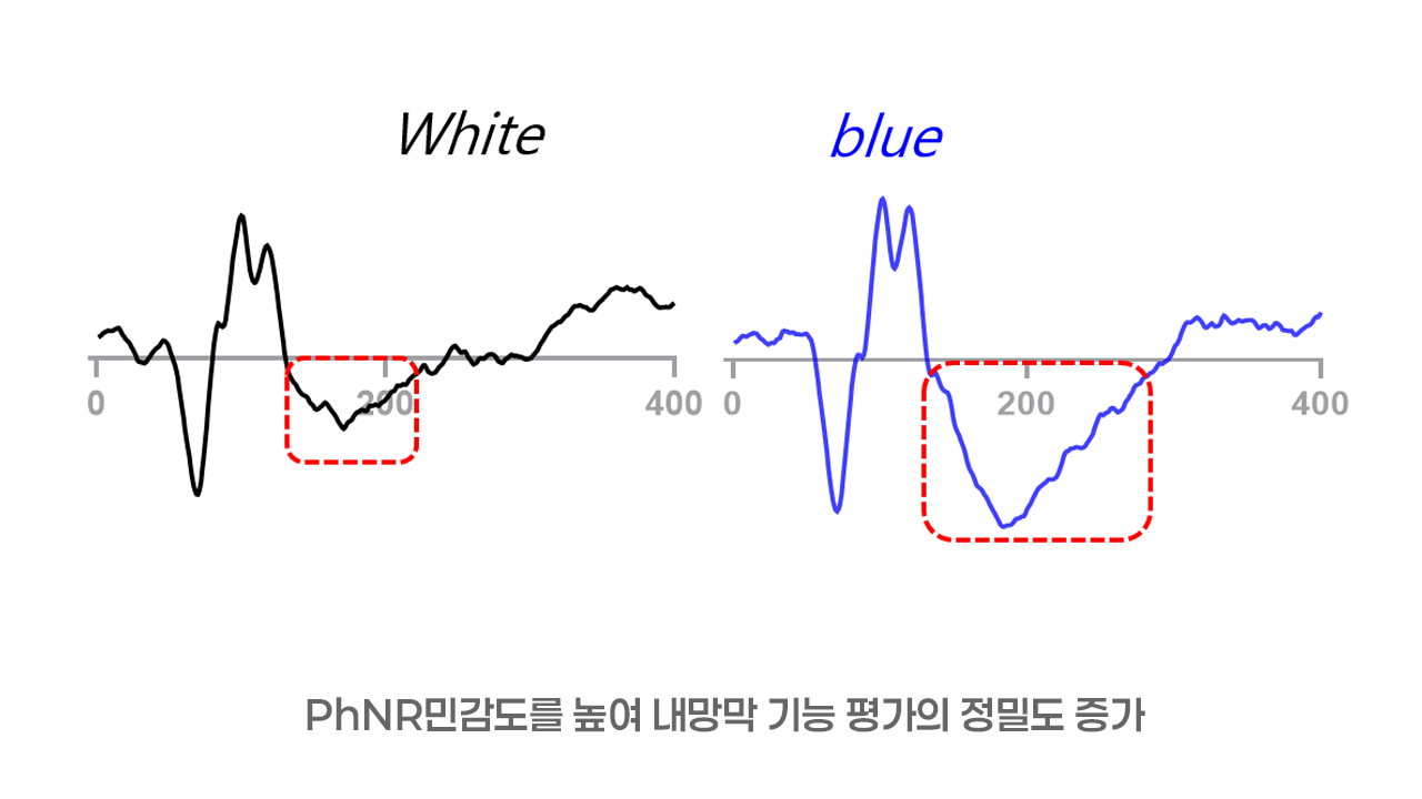
- PhNR, pattern ERG
- Immunohistochemistry
- Quantification of ganglion cell loss
Retinal Detachment model
- Image-guided OCT / funduscopy
- ERG
- H&E Staining
- Immunohistochemistry
Glaucoma model
- Image-guided OCT / funduscopy
- PhNR, pattern ERG
- Immunohistochemistry
- Quantification of ganglion cell loss
Scopolamine induced dry eye mouse model
BAC induced dry eye rabbit model
Presbyopia (Lens Elasticity study)
Ocular PK/PD
System
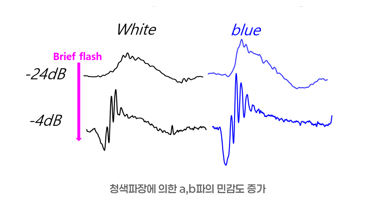
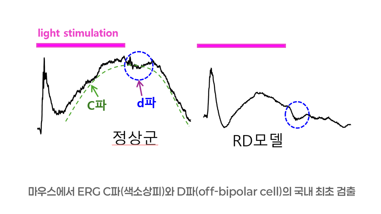
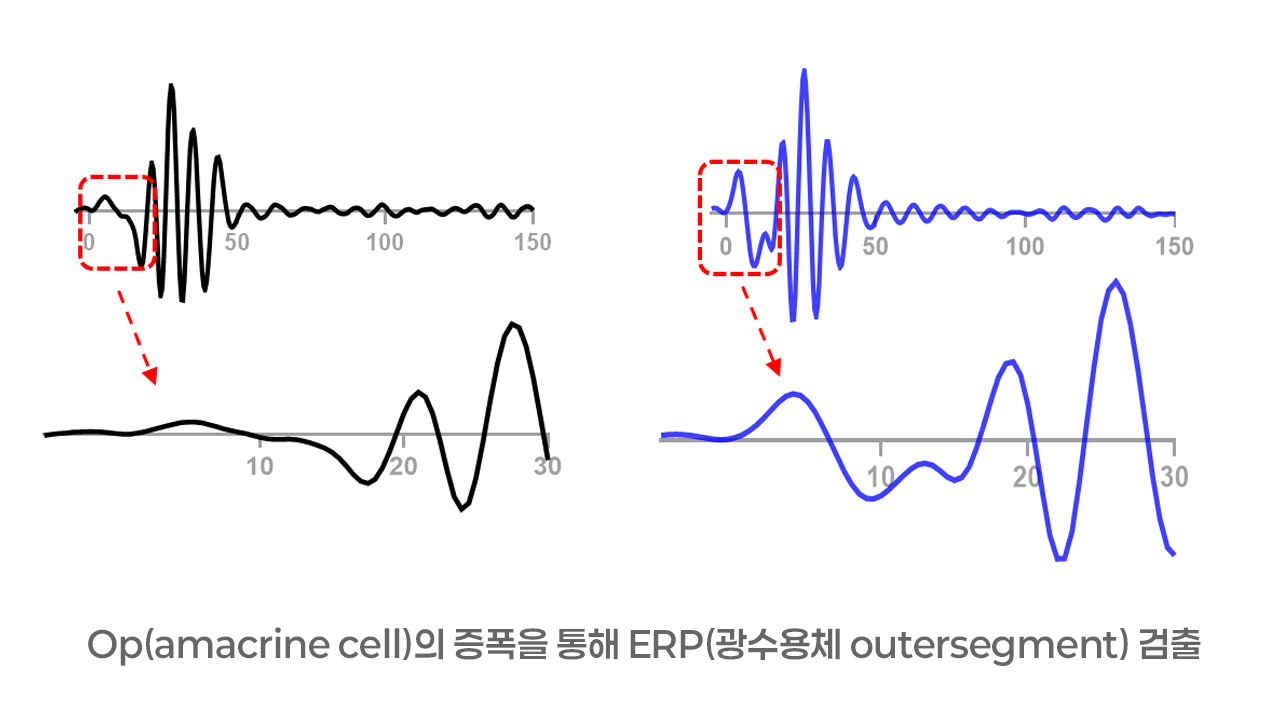
A novel analysis of ERG responses optimized for mouse retinal function
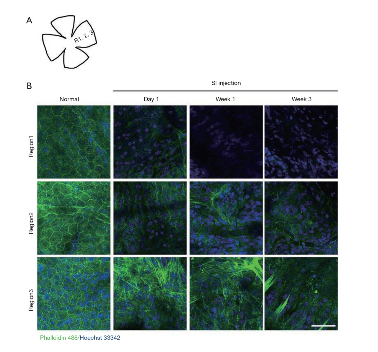
A whole-mount technology uniquely enabled by our proprietary core innovation
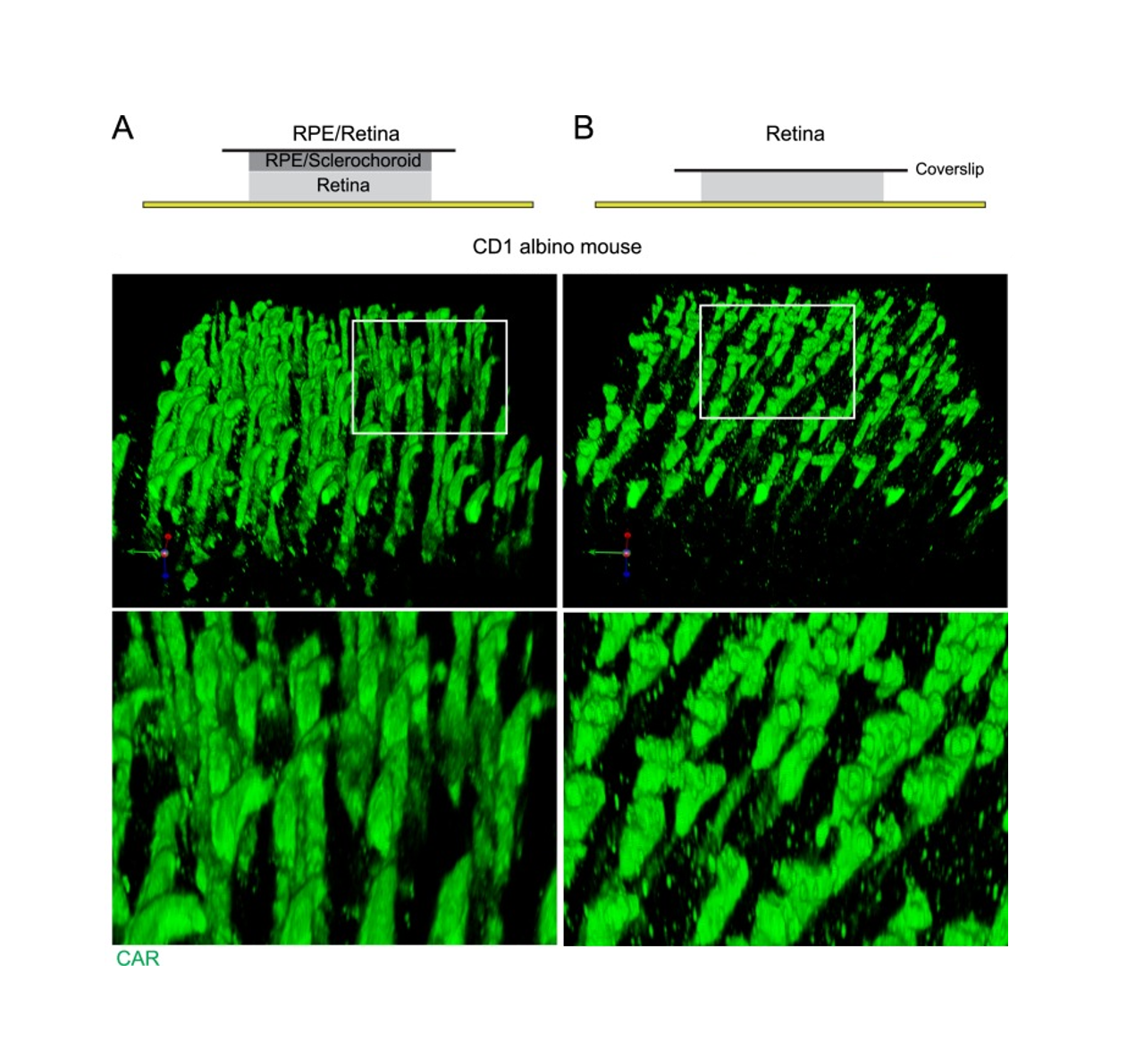
Histology evaluation in non-separate sclerochoroid/retina pigment epithelium/retina whole mounts from C57/BL6 mouse
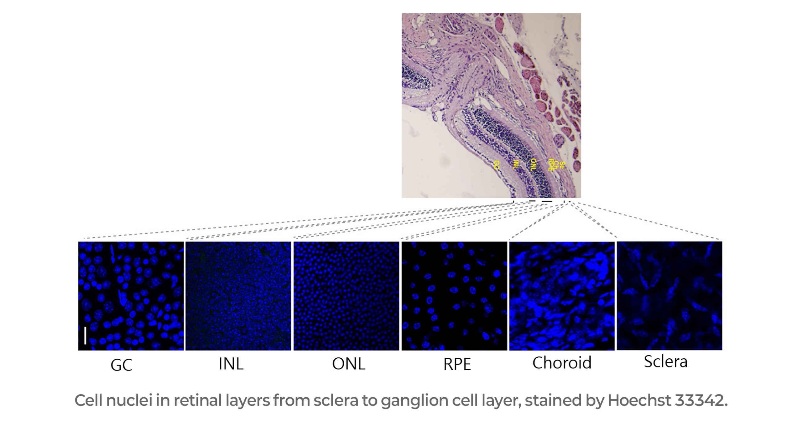
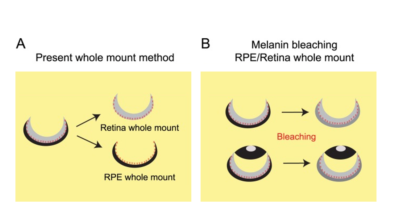
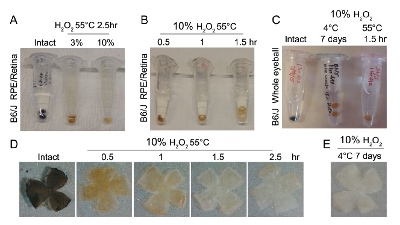
A new method of pigment bleaching RPE / Retina whole mount to observe intact subretinal space